Abstract
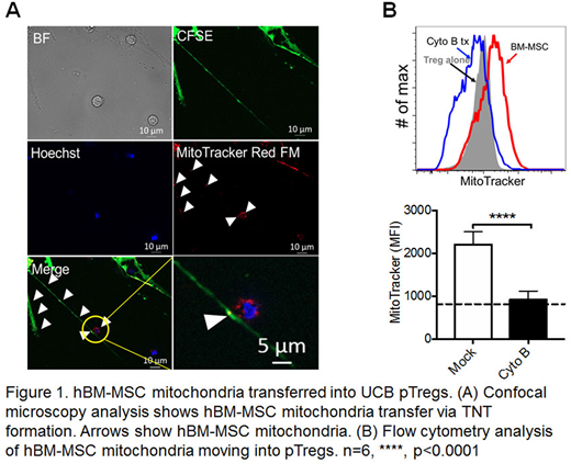
Tumor growth factor β (TGF-β)-induced peripheral regulatory T cells (pTreg) are a promising therapeutic cell source that exhibit Foxp3 expression and suppressive functions similar to natural regulatory T cells. Nonetheless, their clinical potential is limited by the instability of Foxp3 expression and T cell exhaustion that occurs during ex vivo expansion. We postulated that mesenchymal stromal cells (MSCs) could enhance the number, function and Foxp3 expression stability of pTregs during IL-2 driven 21 day expansion due to their diverse immunomodulatory properties. In this study, we observed that use of a human bone marrow mesenchymal stromal cells (hBM-MSC) platform significantly enhanced the number of pTreg during IL-2 driven 21 day ex vivo expansion vs. standard suspension culture condition (MSC platform: 80.2 x 10⁶ vs. IL2/media: 39.3 x 10⁶, n=6; p<0.01). Also the number of pTreg expressing a naive phenotype (CD4+CD45RA+ and CD4+CD62L+ ) were significantly increased (CD45RA+; MSC platform: 74.4 ± 1.6 x 10⁶ vs. IL2/media: 45.9 ± 2.9 x 10⁶, n=6, p<0.001; CD62L+; MSC platform: 79.1 ± 1.3 x 10⁶ vs. IL2/media: 54.5 ± 2.1 x 10⁶, n=6, p<0.001), as well as stability of Foxp3 expression (IL-2/media: 88.2 ± 1.7% vs. MSC platform: 96.2 ± 1.1%, n=7; p<0.05). In addition, pTreg suppressive function was noted to be more potent during 21 day IL-2 driven ex vivo expansion compared to standard IL-2/media culture condition (MSC platform: 79% vs. media: 35% inhibition of T cell proliferation in 10:1 ratio, n=6; p<0.01). pTreg expanded over a hBM-MSC platform exhibited higher surface CD25, CTLA-4, and ICOS MFI expression (CD25; MSC platform: 1410 vs. Media: 774; p<0.001, CTLA-4; MSC platform: 1084 vs. Media: 318; p<0.001, ICOS; MSC platform: 4386 vs. Media: 2641, p<0.01, n=6). Notably, hBM-MSC enhancement of pTreg ex vivo expansion requires direct cell-cell contact, as Foxp3 expression in pTreg was not enhanced by hBM-MSC conditioned media (CM:73.4 ± 6.8% vs. MSC platform: 96.2 ± 1.0%, p<0.001; and IL2/media: 88.8 ± 1.6% vs. MSC platform: 96.2 ± 1.0%, p<0.01) nor in a trans-well culture experiments (Transwell: 83.4 ± 2.5% vs. IL2/media: 88.8 ± 1.6%; and Transwell: 83.4 ± 2.5% vs. MSC platform: 96.2 ± 1.0%, p<0.01). Importantly, optical sectioning microscopy and flow cytometry revealed that hBM-MSC supports Treg number and function via direct contact-dependent mitochondrial transfer (Figure 1A-B). Cytochalasin B treatment blocked mitochondrial transfer, suggesting that tunneling nanotubes (TNT) facilitate mitochondrial transfer from hBM-MSC to pTreg during IL-2 driven ex vivo expansion (Mock: 2208 ± 122.1 vs. Cyto B: 923.8 ± 89 MFI, n=6, p<0.0001). Moreover, the quantity of ATP (n=6; p<0.01) mitochondrial potential of pTreg (MSC platform: 9010 ± 224.5 vs. media: 7316 ± 122.7 MFI, n=6; p<0.01) were significantly enhanced in pTreg during IL-2 driven ex vivo expansion over a hBM-MSC platform. Taken together, hBM-MSC significantly improves the number, maturation, and function of pTreg during 21 day IL-2 driven ex vivo expansion. We have identified one key mechanism of action of hBM-MSC underlying these favorable effects on pTreg during ex vivo expansion to be mitochondrial transfer via TNT. Notably, these studies identify a novel role of hBM-MSC to overcome current limitations in IL-2/media suspension culture conditions including T cell senescence, and loss of Foxp3 expression.
No relevant conflicts of interest to declare.
Author notes
Asterisk with author names denotes non-ASH members.


This feature is available to Subscribers Only
Sign In or Create an Account Close Modal